






Molecular Neuro-Oncology Research
We study how pediatric brain tumors invade the brain and interact with their neural microenvironment, and use these insights to develop more precise, less toxic therapies for children.
Precise therapies for pediatric brain tumors
Understanding tumor invasion and its interaction with the neural microenvironment to develop safer treatments for children.
Malignant pediatric brain tumors such as medulloblastoma remain a leading cause of cancer-related morbidity and mortality in children, despite advances in surgery, radiation, and chemotherapy. Survivors often live with lifelong side effects of these intensive treatments, and some patients continue to die from metastatic disease. Mechanism-based treatments are urgently needed to provide patients with more precise and less toxic therapies.
Brain tumors grow by invading healthy brain tissue and can spread to distant sites within the central nervous system. Our research investigates the molecular mechanisms that allow tumor cells to invade the brain, interact with the neural tumor microenvironment (nTME), and disseminate to metastatic sites. Building on these insights, we develop and functionally validate novel small-molecule strategies that disrupt the mechanisms driving tumor–tissue interaction and invasion.
Current Projects
The tumor in its tissue context: mechanisms and consequences of tumor–tissue interaction
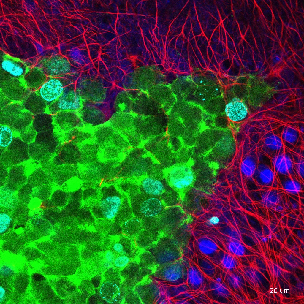
Using cerebellar slice cultures and advanced imaging, we investigate how medulloblastoma cells and their neural microenvironment reshape each other, and how this interplay can be targeted to suppress tumor growth and invasion.
Medulloblastoma (MB) arises in the cerebellum, from where it can invade locally and spread to the meninges of the brain and spinal cord. The neural tumor microenvironment (nTME) of the cerebellum, with its unique cellular, structural, and chemical composition, shapes how MB cells behave. In turn, the growing tumor continuously remodels the nTME through direct contact and by releasing proteins, lipids, nucleic acids, and metabolites. This reciprocal interplay, and its consequences for tumor growth and progression, remains incompletely understood.
Using organotypic cerebellar slice cultures and advanced imaging technologies, we investigate how the growth and invasion of Sonic Hedgehog (SHH) MB cells modify the nTME, and how these changes in turn control tumor cell behavior.
Our primary goal is to identify the molecular mediators of this dynamic reciprocity and define their role in tumor growth and tissue invasion. Our secondary goal is to explore strategies that target these mediators to suppress MB growth and invasion.
Extracellular vesicles in medulloblastoma
We study how medulloblastoma cells use extracellular vesicles to communicate with surrounding brain tissue, aiming to find druggable regulators of vesicle release and new biomarkers in blood or cerebrospinal fluid.
Cells communicate in part by releasing extracellular vesicles (EVs), which carry proteins, RNA, and metabolites from the cell of origin. How medulloblastoma (MB)-derived EVs affect cells in the surrounding cerebellar tissue remains poorly understood.
Using biochemical, proteomic, and sensor-based methods, we determine the composition, regulation, and release–uptake dynamics of MB-derived EVs, identify their target cells, and characterize the transcriptomic changes they induce in those cells.
Our primary goal is to identify druggable modulators of EV biogenesis and release in tumor cells. Our secondary goal is to establish a rational basis for diagnostic or predictive analysis of EV content in blood or cerebrospinal fluid.
Expression, function, and targeting of tumor-associated B7-H3
We investigate the role of the surface molecule B7-H3 in medulloblastoma and develop B7-H3-directed nanobodies to deliver diagnostic and therapeutic payloads to tumor cells.
The tumor-associated surface molecule B7-H3 (CD276) is consistently expressed across many primary pediatric brain tumors, including medulloblastoma (MB), and is associated with poor outcomes. In recent years, B7-H3 has emerged as one of the most compelling cell-surface targets in pediatric CNS malignancies. Yet little is known about its biology in MB: how B7-H3 shapes tumor cell behavior and tissue responses, and how B7-H3-targeted therapies affect signaling between the tumor and its microenvironment.
Using live-cell imaging, gain- and loss-of-function studies, and B7-H3-directed nanobodies, we characterize B7-H3 function in the cerebellar tissue context and explore nanobodies as a strategy to deliver diagnostic and therapeutic payloads to tumor cells.
Image-based morphological and functional analysis of tumor cell behavior at the single-cell level
We combine live-cell imaging, fluorescent sensors, and deep learning to quantify how tumor cells move and invade, and to assess treatment effects at the single-cell level.
We use live- and fixed-cell imaging to analyze single cells, both in isolation and in their tissue context, qualitatively and quantitatively. The lab has developed pipelines for robust quantification of cell dynamics and functions in two- and three-dimensional environments, and uses fluorescent probes and sensors to interrogate specific proteins and measure cellular responses to treatment.
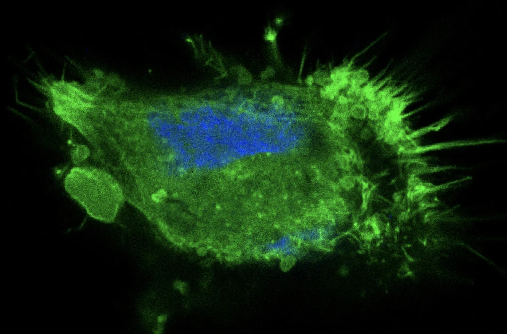
Conventional image analysis cannot capture the changes in F-actin architecture that define motile, invasive cancer cell behavior. To quantify these phenotype-defining features, we are integrating deep learning into our phenotype analyses. We are developing image classifiers that predict treatment efficacy, and generative models that build an interpretable representation of the phenotypic information in the F-actin cytoskeleton of control and treated cells. We use these tools to assess both new and established compounds in an unbiased way.
SNSF Sinergia Project: Rational Small Molecule Inhibition of Dissemination (RaSMID) in pediatric brain tumors
In this SNSF Sinergia project, we designed and validated more than 200 novel small molecules, identified promising lead compounds and new druggable vulnerabilities in medulloblastoma, and established new methods for phenotype-based compound analysis.
A major obstacle to developing anti-metastatic treatments is the lack of drugs that precisely target the biological mechanisms driving tumor progression. Discovering effective and safe small-molecule inhibitors requires advanced computational (in silico) screening, robust functional validation technologies, and physiologically relevant models to rule out toxicity and determine how compounds affect tumor cell biology.
In this project, we designed, synthesized, and functionally validated more than 200 novel compounds. We identified a shortlist of compounds with the desired properties and uncovered new druggable vulnerabilities in medulloblastoma (MB). The project also produced new methods for phenotype-based analysis of bioactive compounds, laying important groundwork for effective and safe treatments for children with MB.
Collaborators
- Prof. Gisbert Schneider, ETH Zürich, Computer-Assisted Drug Design
- Prof. Stephan Neuhauss, UZH, Neuhauss Group
Innosuisse project: A microfluidic platform to study cellular dynamics from the network to the subcellular scale in ex vivo tissue slices
We developed PHIROS, a microfluidic platform for multi-day, high-resolution imaging of brain tissue slices, and used it to reveal how medulloblastoma cells interact with astrocytes and invade surrounding tissue.
Replicating the cytoarchitecture and cellular heterogeneity of the brain in vitro remains challenging. In this project, we developed PHIROS, a microfluidic platform that supports both the culture of organotypic tissue slices and their extended imaging at subcellular resolution.
Using PHIROS, we characterized medulloblastoma (MB) cell behavior in a physiological tumor microenvironment. We observed dynamic, F-actin-driven interactions between tumor cells and tissue-resident astrocytes, and found that the immune checkpoint molecule B7-H3 localizes to the leading edge of invading tumor cells. We also quantified mitochondrial transfer through tubular connections between tumor cells and astrocytes. Together, these findings show that PHIROS is a versatile system for mechanistic studies in a tissue context, combining controlled compound exposure with high-resolution imaging in physiologically relevant settings.
Collaborators
- Dr. Mario Modena, ETH Zurich, Department of Biosystems Science and Engineering
- Dr. Jana Petr, ETH Zurich, Department of Biosystems Science and Engineering
- Prof. Andreas Hierlemann, ETH Zurich, Department of Biosystems Science and Engineering